
Laboratory of
Ocular Biomechanics
University of Pittsburgh
Copyright Notice: The publishers hold the copyright of these articles. The PDFs are provided here to ensure rapid dissemination of scholarly work. It is understood that you will use them only in a manner consistent with the fair-use provisions of the relevant copyright laws. You may not distribute them or use them for any commercial enterprise.
bioRxiv Notice: Papers in biorXiv have not completed peer-review yet.
Publications
See publications of Dr. Sigal in PubMed, Google Scholar and Research Gate.


loading publications
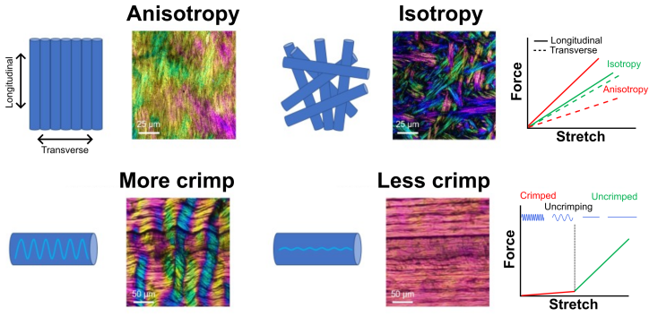
- Collagen microarchitecture from polarized light imaging: A biomechanics perspective
- Accepted by Journal of Biomedical Optics December 2025.


- Collagen microarchitecture from polarized light imaging: A biomechanics perspective
- Accepted by Journal of Biomedical Optics December 2025.
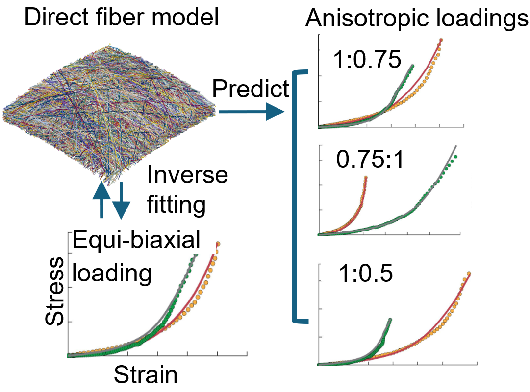
- Capturing sclera anisotropy using direct collagen fiber models; Linking microstructure to macroscopic mechanical properties
- Accepted by Biomechanics and Modeling in Mechanobiology on 23 November 2025. Preprint posted to biorxiv on September 2024.
- Capturing sclera anisotropy using direct collagen fiber models; Linking microstructure to macroscopic mechanical properties
- Accepted by Biomechanics and Modeling in Mechanobiology on 23 November 2025. Preprint posted to biorxiv on September 2024.

- Lamina cribrosa shape in non-human primates is different from that of humans
- Experimental eye research, 261, 110696. https://doi.org/10.1016/j.exer.2025.110696
- Lamina cribrosa shape in non-human primates is different from that of humans
- Experimental eye research, 261, 110696. https://doi.org/10.1016/j.exer.2025.110696
- Selective Deletion of NBCe1 in Reactive Astrocytes Attenuates Ischemic Stroke Brain Damage
- Glia, 73(12), 2386–2406. https://doi.org/10.1002/glia.70075
- Selective Deletion of NBCe1 in Reactive Astrocytes Attenuates Ischemic Stroke Brain Damage
- Glia, 73(12), 2386–2406. https://doi.org/10.1002/glia.70075
- Multiscale analysis of equatorial sclera anisotropy: Revealing discrepancies in fiber orientation and mechanical properties
- Science advances, 11(28), eadp8631. https://doi.org/10.1126/sciadv.adp8631
- Multiscale analysis of equatorial sclera anisotropy: Revealing discrepancies in fiber orientation and mechanical properties
- Science advances, 11(28), eadp8631. https://doi.org/10.1126/sciadv.adp8631

- Pericytes in the optic nerve head
- Prog Retin Eye Res. 2025 May 29:101375. doi: 10.1016/j.preteyeres.2025.101375. PMID: 40449651.
- Pericytes in the optic nerve head
- Prog Retin Eye Res. 2025 May 29:101375. doi: 10.1016/j.preteyeres.2025.101375. PMID: 40449651.

- Lamina cribrosa hypoxia sensitivity to variations of anatomy and vascular factors
- J Biomech Eng. 2025 Aug 1;147(8):081002. doi: 10.1115/1.4068577. PMID: 40314737.
- Lamina cribrosa hypoxia sensitivity to variations of anatomy and vascular factors
- J Biomech Eng. 2025 Aug 1;147(8):081002. doi: 10.1115/1.4068577. PMID: 40314737.
- [Video]

- Impact of elevated IOP on lamina cribrosa oxygenation; A combined experimental-computational study on monkeys

- Morphological comparison of astrocytes in the lamina cribrosa and glial lamina
- Invest Ophthalmol Vis Sci. 2025 Mar 3;66(3):1. doi: 10.1167/iovs.66.3.1. PMID: 40029245; PMCID: PMC11887932..
- Morphological comparison of astrocytes in the lamina cribrosa and glial lamina
- Invest Ophthalmol Vis Sci. 2025 Mar 3;66(3):1. doi: 10.1167/iovs.66.3.1. PMID: 40029245; PMCID: PMC11887932.
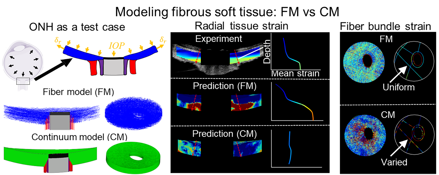
- Comparing continuum and direct fiber models of soft tissues. An ocular biomechanics example reveals that continuum models may artificially disrupt the strains at both the tissue and fiber levels
- Comparing continuum and direct fiber models of soft tissues. An ocular biomechanics example reveals that continuum models may artificially disrupt the strains at both the tissue and fiber levels
- Acta Biomater. 2024 Dec;190:317-328. doi: 10.1016/j.actbio.2024.10.019. PMID: 39424020.
- Proposing a methodology for axon-centric analysis of IOP-induced mechanical insult
- Invest Ophthalmol Vis Sci. 2024 Nov 4;65(13):1. doi: 10.1167/iovs.65.13.1. PMID: 39495185; PMCID: PMC11539975.
- Proposing a methodology for axon-centric analysis of IOP-induced mechanical insult
- Invest Ophthalmol Vis Sci. 2024 Nov 4;65(13):1. doi: 10.1167/iovs.65.13.1. PMID: 39495185; PMCID: PMC11539975.

- Lamina Cribrosa Insertions Into the Sclera Are Sparser, Narrower, and More Slanted in the Anterior Lamina
- Invest Ophthalmol Vis Sci. 2024;65(4):35. doi:10.1167/iovs.65.4.35
- Lamina Cribrosa Insertions Into the Sclera Are Sparser, Narrower, and More Slanted in the Anterior Lamina
- Invest Ophthalmol Vis Sci. 2024;65(4):35. doi:10.1167/iovs.65.4.35

- The Robust Lamina Cribrosa Vasculature: Perfusion and Oxygenation Under Elevated Intraocular Pressure
- Invest Ophthalmol Vis Sci. 2024;65(5):1. doi:10.1167/iovs.65.5.1
- The Robust Lamina Cribrosa Vasculature: Perfusion and Oxygenation Under Elevated Intraocular Pressure
- Invest Ophthalmol Vis Sci. 2024;65(5):1. doi:10.1167/iovs.65.5.1

- Fibrous finite element modeling of the optic nerve head region
- Acta Biomaterialia 2024;175:123-137. doi:10.1016/j.actbio.2023.12.034.
- Fibrous finite element modeling of the optic nerve head region
- Acta Biomaterialia 2024;175:123-137. doi:10.1016/j.actbio.2023.12.034.

- Direct measurements of collagen fiber recruitment in the posterior pole of the eye
- Acta Biomaterialia 2024 Jan 1:173:135-147. doi: 10.1016/j.actbio.2023.11.013
- Direct measurements of collagen fiber recruitment in the posterior pole of the eye
- Acta Biomaterialia 2024 Jan 1:173:135-147. doi: 10.1016/j.actbio.2023.11.013

- 2D or not 2D? Mapping the in-depth inclination of the collagen fibers of the corneoscleral shell
- Experimental Eye Research 2023 Dec:237:109701. doi: 10.1016/j.exer.2023.109701
- 2D or not 2D? Mapping the in-depth inclination of the collagen fibers of the corneoscleral shell
- Experimental Eye Research 2023 Dec:237:109701. doi: 10.1016/j.exer.2023.109701

- A direct fiber approach to model sclera collagen architecture and biomechanics
- Experimental Eye Research. 2023 Jul:232:109510. doi: 10.1016/j.exer.2023.109510
- A direct fiber approach to model sclera collagen architecture and biomechanics
- Experimental Eye Research. 2023 Jul:232:109510. doi: 10.1016/j.exer.2023.109510

- Predatory Bacteria can Reduce Pseudomonas aeruginosa Induced Corneal Perforation and Proliferation in a Rabbit Keratitis Model
- Ocul Surf. 2023 Apr:28:254-261. doi: 10.1016/j.jtos.2023.05.002
- Predatory Bacteria can Reduce Pseudomonas aeruginosa Induced Corneal Perforation and Proliferation in a Rabbit Keratitis Model
- Ocul Surf. 2023 Apr:28:254-261. doi: 10.1016/j.jtos.2023.05.002

- Instant polarized light microscopy pi (IPOLπ) for quantitative imaging of collagen architecture and dynamics in ocular tissues
- Optics and Lasers in Engineering, July 2023, PMID: 36778384 doi: j.optlaseng.2023.107594
- Instant polarized light microscopy pi (IPOLπ) for quantitative imaging of collagen architecture and dynamics in ocular tissues
- Optics and Lasers in Engineering, July 2023, PMID: 36778384 doi: j.optlaseng.2023.107594

- Individual astrocyte morphology in the collagenous lamina cribrosa revealed by multicolor DiOlistic labeling
- Experimental Eye Research, May 2023, PMID: 36965593 doi:2022.12.24.520184
- Individual astrocyte morphology in the collagenous lamina cribrosa revealed by multicolor DiOlistic labeling
- Experimental Eye Research, May 2023, PMID: 36965593 doi:2022.12.24.520184

- Who bears the load? IOP-induced collagen fiber recruitment over the corneoscleral shell
- Experimental Eye Research, May 2023, PMID: 36935071 doi:j.exer.2023.109446.
- Who bears the load? IOP-induced collagen fiber recruitment over the corneoscleral shell
- Experimental Eye Research, May 2023, PMID: 36935071 doi:j.exer.2023.109446.

- Stretch-induced uncrimping of equatorial sclera collagen bundle
- ASME Journal of Biomechanical Engineering, Nov 2022, 2022.09.13.507860
- Stretch-induced uncrimping of equatorial sclera collagen bundle
- ASME Journal of Biomechanical Engineering, Nov 2022, 2022.09.13.507860

- Comparing acute IOP-induced lamina cribrosa deformations pre-mortem and post-mortem
- Translational Vision Science & Technology, Oct 2022, 2022.09.18.508448
- Comparing acute IOP-induced lamina cribrosa deformations pre-mortem and post-mortem
- Translational Vision Science & Technology, Oct 2022, 2022.09.18.508448

- Evidence of an Annexin A4 mediated plasma membrane repair response to biomechanical strain associated with glaucoma pathogenesis
- Evidence of an Annexin A4 mediated plasma membrane repair response to biomechanical strain associated with glaucoma pathogenesis
- Journal of Cellular Physiology, 2022 Sep;237(9):3687-3702, Epub July 21. doi: 10.1002/jcp.30834. PMID: 35862065.


- A high-accuracy and high-efficiency digital volume correlation method to characterize in-vivo optic nerve head biomechanics from optical coherence tomography
- A high-accuracy and high-efficiency digital volume correlation method to characterize in-vivo optic nerve head biomechanics from optical coherence tomography
- Acta Biomaterialia, 2022;143:72-86. April 2022, doi:10.1016/j.actbio.2022.02.021. PMID 35196556.

- A workflow for 3D reconstruction and quantification of the monkey optic nerve head vascular network
- ASME Journal of Biomechanical Engineering, 2022;144(6):061006. June 2022, doi:10.1115/1.4054056
- A workflow for 3D reconstruction and quantification of the monkey optic nerve head vascular network
- ASME Journal of Biomechanical Engineering, 2022;144(6):061006. June 2022, doi:10.1115/1.4054056

- Real-time imaging of optic nerve head collagen microstructure and biomechanics using instant polarized light microscopy
- Experimental eye research, 217, 109867, April 2022.
- Real-time imaging of optic nerve head collagen microstructure and biomechanics using instant polarized light microscopy
- Experimental eye research, 217, 109867, April 2022.

- Lamina cribrosa vessel and collagen beam networks are distinct
- Experimental eye research, 215, 108916, December 2021.
- Lamina cribrosa vessel and collagen beam networks are distinct
- Experimental eye research, 215, 108916, December 2021.

- Interplay between intraocular and intracranial pressure effects on the optic nerve head in vivo
- Experimental eye research, 213, 108809, December 2021.
- Interplay between intraocular and intracranial pressure effects on the optic nerve head in vivo
- Experimental eye research, 213, 108809, December 2021.

- Instant polarized light microscopy for imaging collagen microarchitecture and dynamics
- Journal of Biophotonics, 14(2), e202000326, February 2021.
- Instant polarized light microscopy for imaging collagen microarchitecture and dynamics
- Journal of Biophotonics, 14(2), e202000326, February 2021.

- So-called lamina cribrosa defects may mitigate IOP-induced neural tissue insult
- Investigative Ophthalmology and Visual Science, 61(15), November 2020.
- (* Authors contributed equally to this manuscript)
- So-called lamina cribrosa defects may mitigate IOP-induced neural tissue insult
- Investigative Ophthalmology and Visual Science, 61(15), November 2020.
- (* Authors contributed equally to this manuscript)

- Lamina cribrosa capillaries straighten as intraocular pressure increases
- Investigative Ophthalmology and Visual Science, 61(2), October 2020.
- Lamina cribrosa capillaries straighten as intraocular pressure increases
- Investigative Ophthalmology and Visual Science, 61(2), October 2020.

- Role of radially aligned scleral collagen fibers in optic nerve head biomechanics
- Experimental eye research, 199, 108188, October 2020.
- Role of radially aligned scleral collagen fibers in optic nerve head biomechanics
- Experimental eye research, 199, 108188, October 2020.

- Collagen fiber interweaving is central to sclera stiffness
- Acta Biomaterialia, 113, 429-437, September 2020.
- Collagen fiber interweaving is central to sclera stiffness
- Acta Biomaterialia, 113, 429-437, September 2020.

- Connective tissue remodeling in myopia and its potential role in increasing risk of glaucoma
- Current Opinion in Biomedical Engineering, 15, 40-50, September 2020.
- Connective tissue remodeling in myopia and its potential role in increasing risk of glaucoma
- Current Opinion in Biomedical Engineering, 15, 40-50, September 2020.

- Effect of iStent Trabecular Micro-Bypass device on outflow system morphology
- Modeling and Artificial Intelligence in Ophthalmology, 2(4), 44-54, June 2020.

- Effect of iStent Trabecular Micro-Bypass device on outflow system morphology
- Modeling and Artificial Intelligence in Ophthalmology, 2(4), 44-54, June 2020.

- Test-retest reproducibility of atomic force microscopy measurements of human trabecular meshwork stiffness
- Modeling and Artificial Intelligence in Ophthalmology, 2(4), 34-43, June 2020.
- Test-retest reproducibility of atomic force microscopy measurements of human trabecular meshwork stiffness
- Modeling and Artificial Intelligence in Ophthalmology, 2(4), 34-43, June 2020.

- Instant polarized light microscopy for real-time wide-field visualization of collagen architecture
- Proceeding of SPIE, Label-free Biomedical Imaging and Sensing (LBIS) 2020, 112510Y, February 2020.

- Instant polarized light microscopy for real-time wide-field visualization of collagen architecture
- Proceeding of SPIE, Label-free Biomedical Imaging and Sensing (LBIS) 2020, 112510Y, February 2020.

- A mesh-free approach to incorporate complex anisotropic and heterogeneous material properties into eye-specific finite element models
- A mesh-free approach to incorporate complex anisotropic and heterogeneous material properties into eye-specific finite element models
- Computer Methods in Applied Mechanics and Engineering, 358, 112654, January 2020.

- Scleral structure and biomechanics
- Progress in Retinal and Eye Research, 74, 100773, January 2020.
- Scleral structure and biomechanics
- Progress in Retinal and Eye Research, 74, 100773, January 2020.

- Structured polarized light microscopy for collagen fiber structure and orientation quantification in thick ocular tissues
- Journal of Biomedical Optics, 23(10), 106001, October 2018.
- Structured polarized light microscopy for collagen fiber structure and orientation quantification in thick ocular tissues
- Journal of Biomedical Optics, 23(10), 106001, October 2018.

- Radial and circumferential collagen fibers are a feature of the peripapillary sclera of human, monkey, pig, cow, goat and sheep
- Investigative Ophthalmology and Visual Science, 59(12), 4763-4774, October 2018.
- Radial and circumferential collagen fibers are a feature of the peripapillary sclera of human, monkey, pig, cow, goat and sheep
- Investigative Ophthalmology and Visual Science, 59(12), 4763-4774, October 2018.

- Thin lamina cribrosa beams have different collagen microstructure than thick beams
- Investigative Ophthalmology and Visual Science, 59(11), 4653-4661, September 2018.
- (* Authors contributed equally to this manuscript)
- Thin lamina cribrosa beams have different collagen microstructure than thick beams
- Investigative Ophthalmology and Visual Science, 59(11), 4653-4661, September 2018.
- (* Authors contributed equally to this manuscript)

- Peripapillary sclera architecture revisited: A tangential fiber model and its biomechanical implications
- Acta Biomaterialia, 79, 113-122, August 2018.
- Peripapillary sclera architecture revisited: A tangential fiber model and its biomechanical implications
- Acta Biomaterialia, 79, 113-122, August 2018.

- Spatial patterns and age-related changes of the collagen crimp in the human cornea and sclera
- Investigative Ophthalmology and Visual Science, 59(7), 2987-2998, June 2018.
- Spatial patterns and age-related changes of the collagen crimp in the human cornea and sclera
- Investigative Ophthalmology and Visual Science, 59(7), 2987-2998, June 2018.

- Tortuous pore path through the glaucomatous lamina cribrosa
- Scientific reports, 8(1), 7281, May 2018.
- Tortuous pore path through the glaucomatous lamina cribrosa
- Scientific reports, 8(1), 7281, May 2018.

- Crimp around the globe: Patterns of collagen crimp across the corneoscleral shell
- Experimental eye research, 172, 159-170, April 2018.
- Crimp around the globe: Patterns of collagen crimp across the corneoscleral shell
- Experimental eye research, 172, 159-170, April 2018.

- Polarized light microscopy for 3D mapping of collagen fiber architecture in ocular tissues
- Journal of Biophotonics, 11(8), e201700356, August 2018.
- Polarized light microscopy for 3D mapping of collagen fiber architecture in ocular tissues
- Journal of Biophotonics, 11(8), e201700356, August 2018.

- Structured polarized light microscopy (SPLM) for mapping collagen fiber orientation of ocular tissues
- Proceeding of SPIE, Micromirror Device Based Systems and Applications X, 105460I, February 2018.
- Structured polarized light microscopy (SPLM) for mapping collagen fiber orientation of ocular tissues
- Proceeding of SPIE, Micromirror Device Based Systems and Applications X, 105460I, February 2018.

- Measuring in-vivo and in-situ ex-vivo the 3D deformation of the lamina cribrosa microstructure under elevated intraocular pressure
- Proceeding of SPIE, Optical Elastography and Tissue Biomechanics V, 1049611, February 2018.
- Measuring in-vivo and in-situ ex-vivo the 3D deformation of the lamina cribrosa microstructure under elevated intraocular pressure
- Proceeding of SPIE, Optical Elastography and Tissue Biomechanics V, 1049611, February 2018.

- Seeing the hidden lamina; Effects of exsanguination on the optic nerve head
- Investigative Ophthalmology and Visual Science, 59, 2564-2575, May 2018.
- Seeing the hidden lamina; Effects of exsanguination on the optic nerve head
- Investigative Ophthalmology and Visual Science, 59, 2564-2575, May 2018.

- Collagen fiber recruitment: a microstructural basis for the nonlinear response of the posterior pole of the eye to increases in intraocular pressure
- Acta Biomaterialia, 72, 295-305, May 2018.
- Collagen fiber recruitment: a microstructural basis for the nonlinear response of the posterior pole of the eye to increases in intraocular pressure
- Acta Biomaterialia, 72, 295-305, May 2018.

- Gaze evoked deformations in optic nerve head drusen: repetitive shearing as a potential factor in the visual and vascular complications
- Ophthalmology, 125(6), 929-937, June 2018.
- Gaze evoked deformations in optic nerve head drusen: repetitive shearing as a potential factor in the visual and vascular complications
- Ophthalmology, 125(6), 929-937, June 2018.

- Cerebrospinal Fluid Pressure; Revisiting Factors Influencing Optic Nerve Head Biomechanics
- Investigative Ophthalmology and Visual Science, 59(1), 154-165, January 2018.
- Cerebrospinal Fluid Pressure; Revisiting Factors Influencing Optic Nerve Head Biomechanics
- Investigative Ophthalmology and Visual Science, 59(1), 154-165, January 2018.

- In-Vivo Effects of Intraocular and Intracranial Pressures on the Lamina Cribrosa Microstructure
- PLoS ONE, 12(11), e0188302, November 2017.
- (* Authors contributed equally to this manuscript)
- In-Vivo Effects of Intraocular and Intracranial Pressures on the Lamina Cribrosa Microstructure
- PLoS ONE, 12(11), e0188302, November 2017.
- (* Authors contributed equally to this manuscript)

- Lamina Cribrosa Pore Shape and Size as Predictors of Neural Tissue Mechanical Insult
- Investigative Ophthalmology and Visual Science, 58(12), 5336-5346, October 2017.
- Lamina Cribrosa Pore Shape and Size as Predictors of Neural Tissue Mechanical Insult
- Investigative Ophthalmology and Visual Science, 58(12), 5336-5346, October 2017.

- Formalin Fixation and Cryosectioning Cause Only Minimal Changes in Shape or Size of Ocular Tissues
- Scientific Reports, 7(1), 12065, September 2017.
- Formalin Fixation and Cryosectioning Cause Only Minimal Changes in Shape or Size of Ocular Tissues
- Scientific Reports, 7(1), 12065, September 2017.

- Location of the Central Retinal Vessel Trunk in the Laminar and Prelaminar Tissue of Healthy and Glaucomatous Eyes
- Scientific Reports, 7(1), 9930, August 2017.
- Location of the Central Retinal Vessel Trunk in the Laminar and Prelaminar Tissue of Healthy and Glaucomatous Eyes
- Scientific Reports, 7(1), 9930, August 2017.

- Effects of collagen microstructure and material properties on the deformation of the neural tissues of the lamina cribrosa
- Acta Biomaterialia, 58, 278-290, August 2017.
- Effects of collagen microstructure and material properties on the deformation of the neural tissues of the lamina cribrosa
- Acta Biomaterialia, 58, 278-290, August 2017.

- Whole-globe biomechanics using high-field MRI
- Experimental Eye Research, 160, 85-95, July 2017.
- Whole-globe biomechanics using high-field MRI
- Experimental Eye Research, 160, 85-95, July 2017.

- Microstructural Crimp of the Lamina Cribrosa and Peripapillary Sclera Collagen Fibers
- Investigative Ophthalmology and Visual Science, 58(9), 3378-3388, July 2017.
- Microstructural Crimp of the Lamina Cribrosa and Peripapillary Sclera Collagen Fibers
- Investigative Ophthalmology and Visual Science, 58(9), 3378-3388, July 2017.

- Thick Prelaminar Tissue Decreases Lamina Cribrosa Visibility
- Investigative Ophthalmology and Visual Science, 58(3), 1751-1757, March 2017.
- Thick Prelaminar Tissue Decreases Lamina Cribrosa Visibility
- Investigative Ophthalmology and Visual Science, 58(3), 1751-1757, March 2017.

- An imaged-based inverse finite element method to determine in-vivo mechanical properties of human trabecular meshwork
- Journal for Modeling in Ophthalmology, 1(3), 100-111, 2017.
- An imaged-based inverse finite element method to determine in-vivo mechanical properties of human trabecular meshwork
- Journal for Modeling in Ophthalmology, 1(3), 100-111, 2017.

- Biomechanical aspects of axonal damage in glaucoma: A brief review
- Experimental eye research, 157, 13-19, April 2017.
- Biomechanical aspects of axonal damage in glaucoma: A brief review
- Experimental eye research, 157, 13-19, April 2017.

- Biological aspects of axonal damage in glaucoma: A brief review
- Experimental eye research, 157, 5-12, April 2017.
- Biological aspects of axonal damage in glaucoma: A brief review
- Experimental eye research, 157, 5-12, April 2017.

- Correlation between cerebral hemodynamic and perfusion pressure changes in non-human primates
- Proceeding of SPIE, Optical Tomography and Spectroscopy of Tissue XII, 100591P, February 2017.
- Correlation between cerebral hemodynamic and perfusion pressure changes in non-human primates
- Proceeding of SPIE, Optical Tomography and Spectroscopy of Tissue XII, 100591P, February 2017.

- Mapping in-vivo optic nerve head strains caused by intraocular and intracranial pressures
- Proceeding of SPIE, Optical Elastography and Tissue Biomechanics IV, 100670B, February 2017.
- Mapping in-vivo optic nerve head strains caused by intraocular and intracranial pressures
- Proceeding of SPIE, Optical Elastography and Tissue Biomechanics IV, 100670B, February 2017.

- Collagen architecture of the posterior pole; high-resolution, wide-field-of-view visualization and analysis using polarized light microscopy
- Investigative Ophthalmology and Visual Science, 58(2), 735744, February 2017.
- Collagen architecture of the posterior pole; high-resolution, wide-field-of-view visualization and analysis using polarized light microscopy
- Investigative Ophthalmology and Visual Science, 58(2), 735744, February 2017.

- Identifying the Palisades of Vogt in Human Ex-vivo Tissue
- The Ocular Surface 14(4), 435-439, August 2016.
- Identifying the Palisades of Vogt in Human Ex-vivo Tissue
- The Ocular Surface 14(4), 435-439, August 2016.

- Non-invasive MRI Assessments of Tissue Microstructures and Macromolecules in the Eye upon Biomechanical or Biochemical Modulation
- Scientific Reports, 6(1), 1-14, August 2016.
- Non-invasive MRI Assessments of Tissue Microstructures and Macromolecules in the Eye upon Biomechanical or Biochemical Modulation
- Scientific Reports, 6(1), 1-14, August 2016.

- Experimental glaucoma causes optic nerve head neural rim tissue compression: a potentially important mechanism of axon injury
- Investigative Ophthalmology and Visual Science, 57(10), 4403-4411, August 2016.
- Experimental glaucoma causes optic nerve head neural rim tissue compression: a potentially important mechanism of axon injury
- Investigative Ophthalmology and Visual Science, 57(10), 4403-4411, August 2016.

- What is a typical optic nerve head?
- Experimental Eye Research, 149, 40-47, June 2016.
- What is a typical optic nerve head?
- Experimental Eye Research, 149, 40-47, June 2016.

- Decreased lamina cribrosa beam thickness and pore diameter relative to distance from the central retinal vessel trunk
- Investigative Ophthalmology and Visual Science, 57(7), 3088-3092, June 2016.
- Decreased lamina cribrosa beam thickness and pore diameter relative to distance from the central retinal vessel trunk
- Investigative Ophthalmology and Visual Science, 57(7), 3088-3092, June 2016.

- Regionally Discrete Aqueous Humor Outflow Quantification Using Fluorescein Canalograms
- PLoS ONE, 11(3), e0151754, March 2016.
- Regionally Discrete Aqueous Humor Outflow Quantification Using Fluorescein Canalograms
- PLoS ONE, 11(3), e0151754, March 2016.

- A Problem of Proportions in OCT-based Morphometry and a Proposed Solution
- Investigative Ophthalmology and Visual Science, 57(2), 484-485, Feburary 2016.
- (Letter to the Editor)
- A Problem of Proportions in OCT-based Morphometry and a Proposed Solution
- Investigative Ophthalmology and Visual Science, 57(2), 484-485, Feburary 2016.
- (Letter to the Editor)

- MAPS – A Magic Angle Positioning System for Enhanced Imaging in High-Field Small-Bore MRI
- Journal of Medical Robotics Research 1(1), 1640004, April 2016.
- [Link]
- MAPS – A Magic Angle Positioning System for Enhanced Imaging in High-Field Small-Bore MRI
- Journal of Medical Robotics Research 1(1), 1640004, April 2016.

- Use and Misuse of Laplace’s Law in Ophthalmology
- Investigative Ophthalmology and Visual Science, 57(1), 236-245, January 2016.
- Use and Misuse of Laplace’s Law in Ophthalmology
- Investigative Ophthalmology and Visual Science, 57(1), 236-245, January 2016.

- Polarization Microscopy for Characterizing Fiber Orientation of Ocular Tissues
- Biomedical Optics Express, 6(12), 4705-4718, December 2015.
- Polarization Microscopy for Characterizing Fiber Orientation of Ocular Tissues
- Biomedical Optics Express, 6(12), 4705-4718, December 2015.

- Translating Ocular Biomechanics into Clinical Practice: Current State and Future Prospects
- Current Eye Research, 40(1), 1-18, Jan 2015.
- Translating Ocular Biomechanics into Clinical Practice: Current State and Future Prospects
- Current Eye Research, 40(1), 1-18, Jan 2015.

- Glaucomatous cupping of the lamina cribrosa: A review of the evidence for active progressive remodeling as a mechanism
- Experimental Eye Research, 93(2), 133-140, August 2011.
- Glaucomatous cupping of the lamina cribrosa: A review of the evidence for active progressive remodeling as a mechanism
- Experimental Eye Research, 93(2), 133-140, August 2011

- In Vivo Evaluation of White Matter Inegrity and Anterograde Transport in Visual Systems After Excitoxic Retinal Injury with Multimodal MRI and OCT
- Investigative Ophthalmology and Visual Science, 56(6), 3788-3800, June 2015.
- In Vivo Evaluation of White Matter Inegrity and Anterograde Transport in Visual Systems After Excitoxic Retinal Injury with Multimodal MRI and OCT
- Investigative Ophthalmology and Visual Science, 56(6), 3788-3800, June 2015.

- Histogram Matching Extends Acceptable Signal Strength Range on Optical Coherence Tomography Images
- Investigative Ophthalmology and Visual Science, 56(6), 3810-3819, June 2015.
- Histogram Matching Extends Acceptable Signal Strength Range on Optical Coherence Tomography Images
- Investigative Ophthalmology and Visual Science, 56(6), 3810-3819, June 2015.

- Trabecular Meshwork Response to Pressure Elevation in the Living Human Eye
- Journal of Visualized Experiments, 20(100), e52611, June 2015.
- Trabecular Meshwork Response to Pressure Elevation in the Living Human Eye
- Journal of Visualized Experiments, 20(100), e52611, June 2015.

- Parameters for lithium treatment are critical in its enhancement of fracture-healing in rodents
- The Journal of bone and joint surgery. American volume, 96(23), 1990-1998, December 2014.
- Parameters for lithium treatment are critical in its enhancement of fracture-healing in rodents
- The Journal of bone and joint surgery. American volume, 96(23), 1990-1998, December 2014.

- Application of Elliptic Fourier Analysis to Describe the Lamina Cribrosa Shape with Age and Intraocular Pressure
- Experimental Eye Research, 128, 1-7, November 2014.
- Application of Elliptic Fourier Analysis to Describe the Lamina Cribrosa Shape with Age and Intraocular Pressure
- Experimental Eye Research, 128, 1-7, November 2014.

- In Vivo Three-Dimensional Characterization of the Healthy Human Lamina Cribrosa with Adaptive Optics Spectral-Domain Optical Coherence Tomography
- Investigative Ophthalmology and Visual Science, 55(10), 6459-6466, October 2014.
- In Vivo Three-Dimensional Characterization of the Healthy Human Lamina Cribrosa with Adaptive Optics Spectral-Domain Optical Coherence Tomography
- Investigative Ophthalmology and Visual Science, 55(10), 6459-6466, October 2014.

- Magic Angle-Enhanced MRI Of Fibrous Microstructures In Sclera And Cornea With And Without Intraocular Pressure Loading
- Investigative Ophthalmology and Visual Science, 55(9), 5662-5672, September 2014.
- (* Authors contributed equally to this manuscript)
- Magic Angle-Enhanced MRI Of Fibrous Microstructures In Sclera And Cornea With And Without Intraocular Pressure Loading
- Investigative Ophthalmology and Visual Science, 55(9), 5662-5672, September 2014.
- (* Authors contributed equally to this manuscript)

- Recent Advances in OCT Imaging of the Lamina Cribrosa
- British Journal of Ophthalmology, 98(Suppl 2), ii34-ii39, July 2014.
- Recent Advances in OCT Imaging of the Lamina Cribrosa
- British Journal of Ophthalmology, 98(Suppl 2), ii34-ii39, June 2014.

- Characterization of Schlemm’s Canal Cross-Sectional Area
- British Journal of Ophthalmology, 98(Suppl 2), ii10-ii14, June 2014.
- Characterization of Schlemm’s Canal Cross-Sectional Area
- British Journal of Ophthalmology, 98(Suppl 2), ii10-ii14, June 2014.

- A Method to Estimate Biomechanics and Mechanical Properties of Optic Nerve Head Tissues From Parameters Measurable Using Optical Coherence Tomography
- IEEE Transactions on Medical Imaging, 33(6), 1381-1389, June 2014.
- A Method to Estimate Biomechanics and Mechanical Properties of Optic Nerve Head Tissues From Parameters Measurable Using Optical Coherence Tomography
- IEEE Transactions on Medical Imaging, 33(6), 1381-1389, June 2014.

- Reproducibility of In-Vivo OCT Measured Three-Dimensional Human Lamina Cribrosa Microarchitecture
- PLoS ONE, 9(4), e95526, April 2014.
- Reproducibility of In-Vivo OCT Measured Three-Dimensional Human Lamina Cribrosa Microarchitecture
- PLoS ONE, 9(4), e95526, April 2014.

- IOP Elevation Reduces Schlemm’s Canal Cross-sectional Area
- Investigative Ophthalmology and Visual Science, 55(3), 1805-1809, March 2014.
- IOP Elevation Reduces Schlemm’s Canal Cross-sectional Area
- Investigative Ophthalmology and Visual Science, 55(3), 1805-1809, March 2014.

- Repeatability of in vivo 3D lamina cribrosa microarchitecture using adaptive optics spectral domain optical coherence tomography
- Biomedical Optics Express, 5(4), 1114-1123, April 2014.
- Repeatability of in vivo 3D lamina cribrosa microarchitecture using adaptive optics spectral domain optical coherence tomography
- Biomedical Optics Express, 5(4), 1114-1123, April 2014.

- Gold Nanorods as a Contrast Agent for Doppler Optical Coherence Tomography
- PLoS One, 9(3), e90690, March 2014.
- Gold Nanorods as a Contrast Agent for Doppler Optical Coherence Tomography
- PLoS One, 9(3), e90690, March 2014.

- Eye-Specific IOP-Induced Displacements and Deformations of Human Lamina Cribrosa
- Investigative Ophthalmology and Visual Science, 55(1), 1-15, January 2014.
- Eye-Specific IOP-Induced Displacements and Deformations of Human Lamina Cribrosa
- Investigative Ophthalmology and Visual Science, 55(1), 1-15, January 2014.

- In-Vivo Lamina Cribrosa Microarchitecture in Healthy and Glaucomatous Eyes as Assessed by Optical Coherence Tomography
- Investigative Ophthalmology and Visual Science, 54(13), 8270-8274, December 2013.
- In-Vivo Lamina Cribrosa Microarchitecture in Healthy and Glaucomatous Eyes as Assessed by Optical Coherence Tomography
- Investigative Ophthalmology and Visual Science, 54(13), 8270-8274, December 2013.

- Automated Lamina Cribrosa Microstructural Segmentation in Optical Coherence Tomography Scans of Healthy and Glaucomatous Eyes
- Biomedical Optics Express, 4(11), 2596-2608, November 2013.
- Automated Lamina Cribrosa Microstructural Segmentation in Optical Coherence Tomography Scans of Healthy and Glaucomatous Eyes
- Biomedical Optics Express, 4(11), 2596-2608, November 2013.

- Signal Normalization Reduces Systematic Measurement Differences Between Spectral Domain Optical Coherence Tomography Devices
- Investigative Ophthalmology and Visual Science, 54(12), 7317-7322, November 2013.
- Signal Normalization Reduces Systematic Measurement Differences Between Spectral Domain Optical Coherence Tomography Devices
- Investigative Ophthalmology and Visual Science, 54(12), 7317-7322, November 2013.

- Individual A-Scan Signal Normalization Between Two Spectral Domain Optical Coherence Tomography Devices
- Investigative Ophthalmology and Visual Science, 54(5),3463-3471, May 2013.
- Individual A-Scan Signal Normalization Between Two Spectral Domain Optical Coherence Tomography Devices
- Investigative Ophthalmology and Visual Science, 54(5), 3463-3471, May 2013.

- High Dynamic Range Imaging Concept-Based Signal Enhancement Method Reduced the Optical Coherence Tomography Measurement Variability
- Investigative Ophthalmology and Visual Science, 54(1), 836-841, January 2013.
- High Dynamic Range Imaging Concept-Based Signal Enhancement Method Reduced the Optical Coherence Tomography Measurement Variability
- Investigative Ophthalmology and Visual Science, 54(1):836-41, Jan 2013. PMID 23299477.

- Human Lamina Cribrosa Insertion and Age
- Investigative Ophthalmology and Visual Science, 53(11), 6780-6789, October 2012.
- Human Lamina Cribrosa Insertion and Age
- Investigative Ophthalmology and Visual Science, 53(11):6780-9, Oct 2012. PMID 22956611.

- Morphometric Analysis of Aqueous Humor Outflow Structures with Spectral Domain Optical Coherence Tomography
- Investigative Ophthalmology and Visual Science, 53(9), 5198-207, September 2012.
- Morphometric Analysis of Aqueous Humor Outflow Structures with Spectral Domain Optical Coherence Tomography
- Invest Opthalmol Vis Sci, 53(9):5198-207, September 2012.

- Visualization of the Conventional Outflow Pathway in the Living Human Eye
- Ophthalmology, 119(8), 1563-1568, August 2012.
- Visualization of the Conventional Outflow Pathway in the Living Human Eye
- Ophthalmology, 119(8), 1563-1568, August 2012.

- A few good responses. Which mechanical effects of IOP on the ONH to study?
- Investigative Ophthalmology and Visual Science, 53(7), 4270-4278, June 2012.
- A few good responses. Which mechanical effects of IOP on the ONH to study?
- Investigative Ophthalmology and Visual Science, 53(7), 4270-4278, June 2012.

- The Optic Nerve Head As A Robust Biomechanical System
- Investigative Ophthalmology and Visual Science, 53(6), 2658-2667, May 2012.
- The Optic Nerve Head As A Robust Biomechanical System
- Investigative Ophthalmology and Visual Science, 53(6), 2658-2667, May 2012.

- Lamina Cribrosa Thickening in Early Glaucoma Predicted by a Microstructure Driven Growth and Remodeling Approach
- Mechanics of Materials, 44, 99-109, January 2012.
- Lamina Cribrosa Thickening in Early Glaucoma Predicted by a Microstructure Driven Growth and Remodeling Approach
- Mechanics of Materials, 44, 99-109, January 2012.

- Effect of Acute Intraocular Pressure Elevation on the Monkey Optic Nerve Head as Detected by Spectral Domain Ocular Coherence Tomography
- Investigative Ophthalmology and Visual Science, 52(12), 9431-9437, December 2011.
- Effect of Acute Intraocular Pressure Elevation on the Monkey Optic Nerve Head as Detected by Spectral Domain Ocular Coherence Tomography
- Investigative Ophthalmology and Visual Science, 52(12), 9431-9437, December 2011.

- IOP-induced lamina cribrosa displacement and scleral canal expansion. Are they independent or related?
- Investigative Ophthalmology and Visual Science, 52(12), 9023-9032. December 2011.
- IOP-induced lamina cribrosa displacement and scleral canal expansion. Are they independent or related?
- Investigative Ophthalmology and Visual Science, 52(12), 9023-9032. December 2011.
- 3D Visualization of Aqueous Outflow Structures In the Living Human Eye
- Experimental eye research, 93(3), 308-315, September 2011.
- Camras Special Issue
- 3D Visualization of Aqueous Outflow Structures In the Living Human Eye
- Experimental eye research, 93(3), 308-315, September 2011.
- Camras Special Issue

- Posterior (outward) migration of the lamina cribrosa and early cupping in monkey experimental glaucoma
- Investigative Ophthalmology and Visual Science, 52(10), 7109-7921, September 2011.
- Posterior (outward) migration of the lamina cribrosa and early cupping in monkey experimental glaucoma
- Investigative Ophthalmology and Visual Science, 52(10), 7109-7921, September 2011.

- An Applet to Estimate the IOP-Induced Stress and Strain Within the Optic Nerve Head
- Investigative Ophthalmology and Visual Science, 52(8), 5497-5506, July 2011.
- An Applet to Estimate the IOP-Induced Stress and Strain Within the Optic Nerve Head
- Investigative Ophthalmology and Visual Science, 52(8), 5497-5506, July 2011.

- Finite Element Modeling of the Human Sclera: Influence on Optic Nerve Head Biomechanics and Connections with Glaucoma
- Experimental Eye Research, 93(1), July 2011.
- Finite Element Modeling of the Human Sclera: Influence on Optic Nerve Head Biomechanics and Connections with Glaucoma
- Experimental Eye Research, 93(1), July 2011.

- IOP-Induced Lamina Cribrosa Displacement and Scleral Canal Expansion: an Analysis of Factor Interactions Using Parameterized Eye-Specific Models
- Investigative Ophthalmology and Visual Science, 52(3), 1896-1907, March 2011.
- IOP-Induced Lamina Cribrosa Displacement and Scleral Canal Expansion: an Analysis of Factor Interactions Using Parameterized Eye-Specific Models
- Investigative Ophthalmology and Visual Science, 52(3), 1896-1907, March 2011.

- Longitudinal Change Detected by Spectral Domain Optical Coherence Tomography in the Glaucomatous Optic Nerve Head and Peripapillary Retina
- Investigative Ophthalmology and Visual Science, 52(3), 1206-1219, March 2011.
- Longitudinal Change Detected by Spectral Domain Optical Coherence Tomography in the Glaucomatous Optic Nerve Head and Peripapillary Retina
- Investigative Ophthalmology and Visual Science, 52(3), 1206-19, March 2011.

- Deformation of the Early Glaucomatous Monkey Optic Nerve Head Connective Tissue Following Acute IOP Elevation Within 3-D Histomorphometric Reconstructions
- Investigative Ophthalmology and Visual Science, 52(1), 345-363, January 2011.
- Deformation of the Early Glaucomatous Monkey Optic Nerve Head Connective Tissue Following Acute IOP Elevation Within 3-D Histomorphometric Reconstructions
- Investigative Ophthalmology and Visual Science, 52(1), 345-363, January 2011.

- Changes in the Biomechanical Response of the Optic Nerve Head in Early Experimental Glaucoma
- Investigative Ophthalmology and Visual Science, 51(11):5675-5684. November 2010, PMID 20538997
- Changes in the Biomechanical Response of the Optic Nerve Head in Early Experimental Glaucoma
- Investigative Ophthalmology and Visual Science, 51(11):5675-5684. November 2010, PMID 20538997

- A Geometric Morphometric Assessment of the Optic Cup in Glaucoma
- Experimental Eye Research, 91(3), 405-414, September 2010.
- A Geometric Morphometric Assessment of the Optic Cup in Glaucoma
- Experimental Eye Research, 91(3), 405-414, September 2010.

- Biomechanical Changes of the Optic Disc
- Ocular Disease: Mechanisms and Management Eds: LA Levin and DM Albert, Saunders (Elsevier), Chapter 20, 704, March 2010.
- ISBN 978-0-7020-2983-7
- Biomechanical Changes of the Optic Disc
- Ocular Disease: Mechanisms and Management Eds: LA Levin and DM Albert, Saunders (Elsevier), Chapter 20, 704, March 2010.
- ISBN 978-0-7020-2983-7

- Dimensions of the Human Sclera: Thickness Measurement and Regional Changes with Axial Length
- Experimental Eye Research, 90(2), 277-284, Feburary 2010.
- Dimensions of the Human Sclera: Thickness Measurement and Regional Changes with Axial Length
- Experimental Eye Research, 90(2), 277-284, Feburary 2010.

- 3D morphometry of the Human Optic Nerve Head
- Experimental Eye Research, 90(1), 70-80, January 2010.
- 3D morphometry of the Human Optic Nerve Head
- Experimental Eye Research, 90(1), 70-80, January 2010.

- Mesh Morphing and Response Surface Analysis: Quantifying Sensitivity of Vertebral Mechanical Behaviour
- Annals of Biomedical Engineering, 38(1), 41-56, January 2010.
- Mesh Morphing and Response Surface Analysis: Quantifying Sensitivity of Vertebral Mechanical Behaviour
- Annals of Biomedical Engineering, 38(1), 41-56, January 2010.

- Morphing Methods to Parameterize Specimen-Specific Finite Element Model Geometries
- Journal of Biomechanics, 43(2), 254-262, Jan 2010.
- Morphing Methods to Parameterize Specimen-Specific Finite Element Model Geometries
- Journal of Biomechanics, 43(2), 254-262, Jan 2010.

- Correlation Between Local Stress and Strain and Lamina Cribrosa Connective Tissue Volume Fraction in Normal Monkey Eyes
- Investigative Ophthalmology and Visual Science, 51(1), 295-307, January 2010.
- Correlation Between Local Stress and Strain and Lamina Cribrosa Connective Tissue Volume Fraction in Normal Monkey Eyes
- Investigative Ophthalmology and Visual Science, 51(1), 295-307, January 2010.

- Deformation of the Normal Monkey Optic Nerve Head Connective Tissues Following Acute IOP Elevation Within 3-D Histomorphometric Reconstructions
- Investigative Ophthalmology and Visual Science, 50(12), 5785-5789. December 2009.
- Deformation of the Normal Monkey Optic Nerve Head Connective Tissues Following Acute IOP Elevation Within 3-D Histomorphometric Reconstructions
- Investigative Ophthalmology and Visual Science, 50(12), 5785-5789. December 2009.

- Interactions Between Geometry and Mechanical Properties on the Optic Nerve Head
- Investigative Ophthalmology and Visual Science, 50(6), 2785-2795, June 2009.
- Interactions Between Geometry and Mechanical Properties on the Optic Nerve Head
- Investigative Ophthalmology and Visual Science, 50(6), 2785-2795, June 2009.

- Biomechanics of the Optic Nerve Head
- Experimental Eye Research, 88(4), 799-807, April 2009.
- Biomechanics of the Optic Nerve Head
- Experimental Eye Research, 88(4), 799-807, April 2009.

- Modeling Individual-Specific Human Optic Nerve Head Biomechanics Part II: Influence of Material Properties
- Biomechanics and Modeling in Mechanobiology, 8(2), 99-109, April 2009.
- Modeling Individual-Specific Human Optic Nerve Head Biomechanics Part II: Influence of Material Properties
- Biomechanics and Modeling in Mechanobiology, 8(2), 99-109, April 2009.

- Modeling Individual-Specific Human Optic Nerve Head Biomechanics Part I: IOP-Induced Deformations and Influence of Geometry
- Biomechanics and Modeling in Mechanobiology, 8(2), 85-98, April 2009.
- Modeling Individual-Specific Human Optic Nerve Head Biomechanics Part I: IOP-Induced Deformations and Influence of Geometry
- Biomech Model Mechanobiol, 8(2), 85-98, Apr 2009.

- Mesh-Morphing Algorithms for Specimen-Specific Finite Element Modeling
- Journal of Biomechanics, 41(7), 1381-1389, April 2008.
- Mesh-Morphing Algorithms for Specimen-Specific Finite Element Modeling
- Journal of Biomechanics, 41(7), 1381-1389, April 2008.

- Predicted Extension, Compression and Shearing of Optic Nerve Head Tissues
- Experimental Eye Research, 85(3), 312-322, September 2007.
- Predicted Extension, Compression and Shearing of Optic Nerve Head Tissues
- Experimental Eye Research, 85(3), 312-322, September 2007.

- Factors Influencing Human Optic Nerve Head Biomechanics
- Investigative Ophthalmology and Visual Science, 46(11), 4189-4199, November 2005.
- Factors Influencing Human Optic Nerve Head Biomechanics
- Investigative Ophthalmology and Visual Science, 46(11), 4189-4199, November 2005.

- Reconstruction of Human Optic Nerve Heads for Finite Element Modeling
- Technology and Health Care: Official Journal of the European Society for Engineering and Medicine, 13(4), 313-329, August 2005.
- Reconstruction of Human Optic Nerve Heads for Finite Element Modeling
- Technology and Health Care: Official Journal of the European Society for Engineering and Medicine, 13(4), 313-329, August 2005.

- Finite Element Modeling of Optic Nerve Head Biomechanics
- Investigative Ophthalmology and Visual Science, 45(12), 4378-4387, December 2004.
- Finite Element Modeling of Optic Nerve Head Biomechanics
- Investigative Ophthalmology and Visual Science, 45(12), 4378-4387, December 2004.

- Investigation Of The Biomechanical Environment Within The Optic Nerve Head By Finite Element Modelling
- Computational Fluid and Solid Mechanics, 2nd MIT Conference on, Boston, June 17-20, 2003, K.J. Bathe (ed.), pp. 1810-1813, Elsevier (ISBN: 0-08-044046-0), 2003.

- Investigation Of The Biomechanical Environment Within The Optic Nerve Head By Finite Element Modelling
- Computational Fluid and Solid Mechanics, 2nd MIT Conference on, Boston, June 17-20, 2003, K.J. Bathe (ed.), pp. 1810-1813, Elsevier (ISBN: 0-08-044046-0), 2003.

- Chirality in Bare and Passivated Gold Nanoclusters
- Physical Review B, 66(7), 073403, August 2002.

- Chirality in Bare and Passivated Gold Nanoclusters
- Physical Review B, 66(7), 073403, August 2002.

Publications
See publications of Dr. Sigal in PubMed, Google Scholar and Research Gate.
loading publications
- Collagen microarchitecture from polarized light imaging: A biomechanics perspective
- Accepted by Journal of Biomedical Optics December 2025.
|
|
|
- Capturing sclera anisotropy using direct collagen fiber models; Linking microstructure to macroscopic mechanical properties
- Accepted by Biomechanics and Modeling in Mechanobiology on 23 November 2025. Preprint posted to biorxiv on September 2024.
|
|
|
- Lamina cribrosa shape in non-human primates is different from that of humans
- Experimental eye research, 261, 110696. https://doi.org/10.1016/j.exer.2025.110696
|
|
|
- Selective Deletion of NBCe1 in Reactive Astrocytes Attenuates Ischemic Stroke Brain Damage
- Glia, 73(12), 2386–2406. https://doi.org/10.1002/glia.70075
|
|
|
- Multiscale analysis of equatorial sclera anisotropy: Revealing discrepancies in fiber orientation and mechanical properties
- Science advances, 11(28), eadp8631. https://doi.org/10.1126/sciadv.adp8631
|
|
|
- Pericytes in the optic nerve head
- Prog Retin Eye Res. 2025 May 29:101375. doi: 10.1016/j.preteyeres.2025.101375. PMID: 40449651.
|
|
|
- Lamina cribrosa hypoxia sensitivity to variations of anatomy and vascular factors
- J Biomech Eng. 2025 Aug 1;147(8):081002. doi: 10.1115/1.4068577. PMID: 40314737.
|
|
|
- Impact of elevated IOP on lamina cribrosa oxygenation; A combined experimental-computational study on monkeys
|
|
- Morphological comparison of astrocytes in the lamina cribrosa and glial lamina
- Invest Ophthalmol Vis Sci. 2025 Mar 3;66(3):1. doi: 10.1167/iovs.66.3.1. PMID: 40029245; PMCID: PMC11887932..
|
|
|
- Comparing continuum and direct fiber models of soft tissues. An ocular biomechanics example reveals that continuum models may artificially disrupt the strains at both the tissue and fiber levels
|
|
|
- Proposing a methodology for axon-centric analysis of IOP-induced mechanical insult
- Invest Ophthalmol Vis Sci. 2024 Nov 4;65(13):1. doi: 10.1167/iovs.65.13.1. PMID: 39495185; PMCID: PMC11539975.
|
|
|
- Lamina Cribrosa Insertions Into the Sclera Are Sparser, Narrower, and More Slanted in the Anterior Lamina
- Invest Ophthalmol Vis Sci. 2024;65(4):35. doi:10.1167/iovs.65.4.35
|
|
|
- The Robust Lamina Cribrosa Vasculature: Perfusion and Oxygenation Under Elevated Intraocular Pressure
- Invest Ophthalmol Vis Sci. 2024;65(5):1. doi:10.1167/iovs.65.5.1
|
|
|
- Fibrous finite element modeling of the optic nerve head region
- Acta Biomaterialia 2024;175:123-137. doi:10.1016/j.actbio.2023.12.034.
|
|
|
- Direct measurements of collagen fiber recruitment in the posterior pole of the eye
- Acta Biomaterialia 2024 Jan 1:173:135-147. doi: 10.1016/j.actbio.2023.11.013
|
|
|
- 2D or not 2D? Mapping the in-depth inclination of the collagen fibers of the corneoscleral shell
- Experimental Eye Research 2023 Dec:237:109701. doi: 10.1016/j.exer.2023.109701
|
|
|
- A direct fiber approach to model sclera collagen architecture and biomechanics
- Experimental Eye Research. 2023 Jul:232:109510. doi: 10.1016/j.exer.2023.109510
|
|
|
- Predatory Bacteria can Reduce Pseudomonas aeruginosa Induced Corneal Perforation and Proliferation in a Rabbit Keratitis Model
- Ocul Surf. 2023 Apr:28:254-261. doi: 10.1016/j.jtos.2023.05.002
|
|
|
- Instant polarized light microscopy pi (IPOLπ) for quantitative imaging of collagen architecture and dynamics in ocular tissues
- Optics and Lasers in Engineering, July 2023, PMID: 36778384 doi: j.optlaseng.2023.107594
|
|
|
- Individual astrocyte morphology in the collagenous lamina cribrosa revealed by multicolor DiOlistic labeling
- Experimental Eye Research, May 2023, PMID: 36965593 doi:2022.12.24.520184
|
|
|
- Who bears the load? IOP-induced collagen fiber recruitment over the corneoscleral shell
- Experimental Eye Research, May 2023, PMID: 36935071 doi:j.exer.2023.109446.
|
|
|
- Stretch-induced uncrimping of equatorial sclera collagen bundle
- ASME Journal of Biomechanical Engineering, Nov 2022, 2022.09.13.507860
|
|
|
- Comparing acute IOP-induced lamina cribrosa deformations pre-mortem and post-mortem
- Translational Vision Science & Technology, Oct 2022, 2022.09.18.508448
|
|
|
- Evidence of an Annexin A4 mediated plasma membrane repair response to biomechanical strain associated with glaucoma pathogenesis
|
|
|
|
|
- A high-accuracy and high-efficiency digital volume correlation method to characterize in-vivo optic nerve head biomechanics from optical coherence tomography
|
|
|
- A workflow for 3D reconstruction and quantification of the monkey optic nerve head vascular network
- ASME Journal of Biomechanical Engineering, 2022;144(6):061006. June 2022, doi:10.1115/1.4054056
|
|
|
- Real-time imaging of optic nerve head collagen microstructure and biomechanics using instant polarized light microscopy
- Experimental eye research, 217, 109867, April 2022.
|
|
|
- Lamina cribrosa vessel and collagen beam networks are distinct
- Experimental eye research, 215, 108916, December 2021.
|
|
|
- Interplay between intraocular and intracranial pressure effects on the optic nerve head in vivo
- Experimental eye research, 213, 108809, December 2021.
|
|
|
- Instant polarized light microscopy for imaging collagen microarchitecture and dynamics
- Journal of Biophotonics, 14(2), e202000326, February 2021.
|
|
|
- So-called lamina cribrosa defects may mitigate IOP-induced neural tissue insult
- Investigative Ophthalmology and Visual Science, 61(15), November 2020.
- (* Authors contributed equally to this manuscript)
|
|
|
- Lamina cribrosa capillaries straighten as intraocular pressure increases
- Investigative Ophthalmology and Visual Science, 61(2), October 2020.
|
|
|
- Role of radially aligned scleral collagen fibers in optic nerve head biomechanics
- Experimental eye research, 199, 108188, October 2020.
|
|
|
- Collagen fiber interweaving is central to sclera stiffness
- Acta Biomaterialia, 113, 429-437, September 2020.
|
|
|
- Connective tissue remodeling in myopia and its potential role in increasing risk of glaucoma
- Current Opinion in Biomedical Engineering, 15, 40-50, September 2020.
|
|
|
- Effect of iStent Trabecular Micro-Bypass device on outflow system morphology
- Modeling and Artificial Intelligence in Ophthalmology, 2(4), 44-54, June 2020.
|
|
|
- Test-retest reproducibility of atomic force microscopy measurements of human trabecular meshwork stiffness
- Modeling and Artificial Intelligence in Ophthalmology, 2(4), 34-43, June 2020.
|
|
|
- Instant polarized light microscopy for real-time wide-field visualization of collagen architecture
- Proceeding of SPIE, Label-free Biomedical Imaging and Sensing (LBIS) 2020, 112510Y, February 2020.
|
|
|
- A mesh-free approach to incorporate complex anisotropic and heterogeneous material properties into eye-specific finite element models
|
|
|
- Scleral structure and biomechanics
- Progress in Retinal and Eye Research, 74, 100773, January 2020.
|
|
|
- Structured polarized light microscopy for collagen fiber structure and orientation quantification in thick ocular tissues
- Journal of Biomedical Optics, 23(10), 106001, October 2018.
|
|
|
- Radial and circumferential collagen fibers are a feature of the peripapillary sclera of human, monkey, pig, cow, goat and sheep
- Investigative Ophthalmology and Visual Science, 59(12), 4763-4774, October 2018.
|
|
|
- Thin lamina cribrosa beams have different collagen microstructure than thick beams
- Investigative Ophthalmology and Visual Science, 59(11), 4653-4661, September 2018.
- (* Authors contributed equally to this manuscript)
|
|
|
- Peripapillary sclera architecture revisited: A tangential fiber model and its biomechanical implications
- Acta Biomaterialia, 79, 113-122, August 2018.
|
|
|
- Spatial patterns and age-related changes of the collagen crimp in the human cornea and sclera
- Investigative Ophthalmology and Visual Science, 59(7), 2987-2998, June 2018.
|
|
|
- Tortuous pore path through the glaucomatous lamina cribrosa
- Scientific reports, 8(1), 7281, May 2018.
|
|
|
- Crimp around the globe: Patterns of collagen crimp across the corneoscleral shell
- Experimental eye research, 172, 159-170, April 2018.
|
|
|
- Polarized light microscopy for 3D mapping of collagen fiber architecture in ocular tissues
- Journal of Biophotonics, 11(8), e201700356, August 2018.
|
|
|
- Structured polarized light microscopy (SPLM) for mapping collagen fiber orientation of ocular tissues
- Proceeding of SPIE, Micromirror Device Based Systems and Applications X, 105460I, February 2018.
|
|
|
- Measuring in-vivo and in-situ ex-vivo the 3D deformation of the lamina cribrosa microstructure under elevated intraocular pressure
- Proceeding of SPIE, Optical Elastography and Tissue Biomechanics V, 1049611, February 2018.
|
|
|
- Seeing the hidden lamina; Effects of exsanguination on the optic nerve head
- Investigative Ophthalmology and Visual Science, 59, 2564-2575, May 2018.
|
|
|
- Collagen fiber recruitment: a microstructural basis for the nonlinear response of the posterior pole of the eye to increases in intraocular pressure
- Acta Biomaterialia, 72, 295-305, May 2018.
|
|
|
- Gaze evoked deformations in optic nerve head drusen: repetitive shearing as a potential factor in the visual and vascular complications
- Ophthalmology, 125(6), 929-937, June 2018.
|
|
|
- Cerebrospinal Fluid Pressure; Revisiting Factors Influencing Optic Nerve Head Biomechanics
- Investigative Ophthalmology and Visual Science, 59(1), 154-165, January 2018.
|
|
|
- In-Vivo Effects of Intraocular and Intracranial Pressures on the Lamina Cribrosa Microstructure
- PLoS ONE, 12(11), e0188302, November 2017.
- (* Authors contributed equally to this manuscript)
|
|
|
- Lamina Cribrosa Pore Shape and Size as Predictors of Neural Tissue Mechanical Insult
- Investigative Ophthalmology and Visual Science, 58(12), 5336-5346, October 2017.
|
|
|
- Formalin Fixation and Cryosectioning Cause Only Minimal Changes in Shape or Size of Ocular Tissues
- Scientific Reports, 7(1), 12065, September 2017.
|
|
|
- Location of the Central Retinal Vessel Trunk in the Laminar and Prelaminar Tissue of Healthy and Glaucomatous Eyes
- Scientific Reports, 7(1), 9930, August 2017.
|
|
|
- Effects of collagen microstructure and material properties on the deformation of the neural tissues of the lamina cribrosa
- Acta Biomaterialia, 58, 278-290, August 2017.
|
|
|
- Whole-globe biomechanics using high-field MRI
- Experimental Eye Research, 160, 85-95, July 2017.
|
|
|
- Microstructural Crimp of the Lamina Cribrosa and Peripapillary Sclera Collagen Fibers
- Investigative Ophthalmology and Visual Science, 58(9), 3378-3388, July 2017.
|
|
|
- Thick Prelaminar Tissue Decreases Lamina Cribrosa Visibility
- Investigative Ophthalmology and Visual Science, 58(3), 1751-1757, March 2017.
|
|
|
- An imaged-based inverse finite element method to determine in-vivo mechanical properties of human trabecular meshwork
- Journal for Modeling in Ophthalmology, 1(3), 100-111, 2017.
|
|
|
- Biomechanical aspects of axonal damage in glaucoma: A brief review
- Experimental eye research, 157, 13-19, April 2017.
|
|
|
- Biological aspects of axonal damage in glaucoma: A brief review
- Experimental eye research, 157, 5-12, April 2017.
|
|
|
- Correlation between cerebral hemodynamic and perfusion pressure changes in non-human primates
- Proceeding of SPIE, Optical Tomography and Spectroscopy of Tissue XII, 100591P, February 2017.
|
|
|
- Mapping in-vivo optic nerve head strains caused by intraocular and intracranial pressures
- Proceeding of SPIE, Optical Elastography and Tissue Biomechanics IV, 100670B, February 2017.
|
|
|
- Collagen architecture of the posterior pole; high-resolution, wide-field-of-view visualization and analysis using polarized light microscopy
- Investigative Ophthalmology and Visual Science, 58(2), 735744, February 2017.
|
|
|
- Identifying the Palisades of Vogt in Human Ex-vivo Tissue
- The Ocular Surface 14(4), 435-439, August 2016.
|
|
|
- Non-invasive MRI Assessments of Tissue Microstructures and Macromolecules in the Eye upon Biomechanical or Biochemical Modulation
- Scientific Reports, 6(1), 1-14, August 2016.
|
|
|
- Experimental glaucoma causes optic nerve head neural rim tissue compression: a potentially important mechanism of axon injury
- Investigative Ophthalmology and Visual Science, 57(10), 4403-4411, August 2016.
|
|
|
- What is a typical optic nerve head?
- Experimental Eye Research, 149, 40-47, June 2016.
|
|
|
- Decreased lamina cribrosa beam thickness and pore diameter relative to distance from the central retinal vessel trunk
- Investigative Ophthalmology and Visual Science, 57(7), 3088-3092, June 2016.
|
|
|
- Regionally Discrete Aqueous Humor Outflow Quantification Using Fluorescein Canalograms
- PLoS ONE, 11(3), e0151754, March 2016.
|
|
|
- A Problem of Proportions in OCT-based Morphometry and a Proposed Solution
- Investigative Ophthalmology and Visual Science, 57(2), 484-485, Feburary 2016.
- (Letter to the Editor)
|
|
|
- MAPS – A Magic Angle Positioning System for Enhanced Imaging in High-Field Small-Bore MRI
- Journal of Medical Robotics Research 1(1), 1640004, April 2016.
- [Link]
|
|
|
- Use and Misuse of Laplace’s Law in Ophthalmology
- Investigative Ophthalmology and Visual Science, 57(1), 236-245, January 2016.
|
|
|
- Polarization Microscopy for Characterizing Fiber Orientation of Ocular Tissues
- Biomedical Optics Express, 6(12), 4705-4718, December 2015.
|
|
|
- Translating Ocular Biomechanics into Clinical Practice: Current State and Future Prospects
- Current Eye Research, 40(1), 1-18, Jan 2015.
|
|
|
- Glaucomatous cupping of the lamina cribrosa: A review of the evidence for active progressive remodeling as a mechanism
- Experimental Eye Research, 93(2), 133-140, August 2011.
|
|
|
- In Vivo Evaluation of White Matter Inegrity and Anterograde Transport in Visual Systems After Excitoxic Retinal Injury with Multimodal MRI and OCT
- Investigative Ophthalmology and Visual Science, 56(6), 3788-3800, June 2015.
|
|
|
- Histogram Matching Extends Acceptable Signal Strength Range on Optical Coherence Tomography Images
- Investigative Ophthalmology and Visual Science, 56(6), 3810-3819, June 2015.
|
|
|
- Trabecular Meshwork Response to Pressure Elevation in the Living Human Eye
- Journal of Visualized Experiments, 20(100), e52611, June 2015.
|
|
|
- Parameters for lithium treatment are critical in its enhancement of fracture-healing in rodents
- The Journal of bone and joint surgery. American volume, 96(23), 1990-1998, December 2014.
|
|
|
- Application of Elliptic Fourier Analysis to Describe the Lamina Cribrosa Shape with Age and Intraocular Pressure
- Experimental Eye Research, 128, 1-7, November 2014.
|
|
|
- In Vivo Three-Dimensional Characterization of the Healthy Human Lamina Cribrosa with Adaptive Optics Spectral-Domain Optical Coherence Tomography
- Investigative Ophthalmology and Visual Science, 55(10), 6459-6466, October 2014.
|
|
|
- Magic Angle-Enhanced MRI Of Fibrous Microstructures In Sclera And Cornea With And Without Intraocular Pressure Loading
- Investigative Ophthalmology and Visual Science, 55(9), 5662-5672, September 2014.
- (* Authors contributed equally to this manuscript)
|
|
|
- Recent Advances in OCT Imaging of the Lamina Cribrosa
- British Journal of Ophthalmology, 98(Suppl 2), ii34-ii39, July 2014.
|
|
|
- Characterization of Schlemm’s Canal Cross-Sectional Area
- British Journal of Ophthalmology, 98(Suppl 2), ii10-ii14, June 2014.
|
|
|
- A Method to Estimate Biomechanics and Mechanical Properties of Optic Nerve Head Tissues From Parameters Measurable Using Optical Coherence Tomography
- IEEE Transactions on Medical Imaging, 33(6), 1381-1389, June 2014.
|
|
|
- Reproducibility of In-Vivo OCT Measured Three-Dimensional Human Lamina Cribrosa Microarchitecture
- PLoS ONE, 9(4), e95526, April 2014.
|
|
|
- IOP Elevation Reduces Schlemm’s Canal Cross-sectional Area
- Investigative Ophthalmology and Visual Science, 55(3), 1805-1809, March 2014.
|
|
|
- Repeatability of in vivo 3D lamina cribrosa microarchitecture using adaptive optics spectral domain optical coherence tomography
- Biomedical Optics Express, 5(4), 1114-1123, April 2014.
|
|
|
- Gold Nanorods as a Contrast Agent for Doppler Optical Coherence Tomography
- PLoS One, 9(3), e90690, March 2014.
|
|
|
- Eye-Specific IOP-Induced Displacements and Deformations of Human Lamina Cribrosa
- Investigative Ophthalmology and Visual Science, 55(1), 1-15, January 2014.
|
|
|
- In-Vivo Lamina Cribrosa Microarchitecture in Healthy and Glaucomatous Eyes as Assessed by Optical Coherence Tomography
- Investigative Ophthalmology and Visual Science, 54(13), 8270-8274, December 2013.
|
|
|
- Automated Lamina Cribrosa Microstructural Segmentation in Optical Coherence Tomography Scans of Healthy and Glaucomatous Eyes
- Biomedical Optics Express, 4(11), 2596-2608, November 2013.
|
|
|
- Signal Normalization Reduces Systematic Measurement Differences Between Spectral Domain Optical Coherence Tomography Devices
- Investigative Ophthalmology and Visual Science, 54(12), 7317-7322, November 2013.
|
|
|
- Individual A-Scan Signal Normalization Between Two Spectral Domain Optical Coherence Tomography Devices
- Investigative Ophthalmology and Visual Science, 54(5),3463-3471, May 2013.
|
|
|
- High Dynamic Range Imaging Concept-Based Signal Enhancement Method Reduced the Optical Coherence Tomography Measurement Variability
- Investigative Ophthalmology and Visual Science, 54(1), 836-841, January 2013.
|
|
|
- Human Lamina Cribrosa Insertion and Age
- Investigative Ophthalmology and Visual Science, 53(11), 6780-6789, October 2012.
|
|
|
- Morphometric Analysis of Aqueous Humor Outflow Structures with Spectral Domain Optical Coherence Tomography
- Investigative Ophthalmology and Visual Science, 53(9), 5198-207, September 2012.
|
|
|
- Visualization of the Conventional Outflow Pathway in the Living Human Eye
- Ophthalmology, 119(8), 1563-1568, August 2012.
|
|
|
- A few good responses. Which mechanical effects of IOP on the ONH to study?
- Investigative Ophthalmology and Visual Science, 53(7), 4270-4278, June 2012.
|
|
|
- The Optic Nerve Head As A Robust Biomechanical System
- Investigative Ophthalmology and Visual Science, 53(6), 2658-2667, May 2012.
|
|
|
- Lamina Cribrosa Thickening in Early Glaucoma Predicted by a Microstructure Driven Growth and Remodeling Approach
- Mechanics of Materials, 44, 99-109, January 2012.
|
|
|
- Effect of Acute Intraocular Pressure Elevation on the Monkey Optic Nerve Head as Detected by Spectral Domain Ocular Coherence Tomography
- Investigative Ophthalmology and Visual Science, 52(12), 9431-9437, December 2011.
|
|
|
- IOP-induced lamina cribrosa displacement and scleral canal expansion. Are they independent or related?
- Investigative Ophthalmology and Visual Science, 52(12), 9023-9032. December 2011.
|
|
|
- 3D Visualization of Aqueous Outflow Structures In the Living Human Eye
- Experimental eye research, 93(3), 308-315, September 2011.
- Camras Special Issue
|
|
|
- Posterior (outward) migration of the lamina cribrosa and early cupping in monkey experimental glaucoma
- Investigative Ophthalmology and Visual Science, 52(10), 7109-7921, September 2011.
|
|
|
- An Applet to Estimate the IOP-Induced Stress and Strain Within the Optic Nerve Head
- Investigative Ophthalmology and Visual Science, 52(8), 5497-5506, July 2011.
|
|
|
- Finite Element Modeling of the Human Sclera: Influence on Optic Nerve Head Biomechanics and Connections with Glaucoma
- Experimental Eye Research, 93(1), July 2011.
|
|
|
- IOP-Induced Lamina Cribrosa Displacement and Scleral Canal Expansion: an Analysis of Factor Interactions Using Parameterized Eye-Specific Models
- Investigative Ophthalmology and Visual Science, 52(3), 1896-1907, March 2011.
|
|
|
- Longitudinal Change Detected by Spectral Domain Optical Coherence Tomography in the Glaucomatous Optic Nerve Head and Peripapillary Retina
- Investigative Ophthalmology and Visual Science, 52(3), 1206-1219, March 2011.
|
|
|
- Deformation of the Early Glaucomatous Monkey Optic Nerve Head Connective Tissue Following Acute IOP Elevation Within 3-D Histomorphometric Reconstructions
- Investigative Ophthalmology and Visual Science, 52(1), 345-363, January 2011.
|
|
|
- Changes in the Biomechanical Response of the Optic Nerve Head in Early Experimental Glaucoma
- Investigative Ophthalmology and Visual Science, 51(11):5675-5684. November 2010, PMID 20538997
|
|
|
- A Geometric Morphometric Assessment of the Optic Cup in Glaucoma
- Experimental Eye Research, 91(3), 405-414, September 2010.
|
|
|
- Biomechanical Changes of the Optic Disc
- Ocular Disease: Mechanisms and Management Eds: LA Levin and DM Albert, Saunders (Elsevier), Chapter 20, 704, March 2010.
- ISBN 978-0-7020-2983-7
|
|
|
- Dimensions of the Human Sclera: Thickness Measurement and Regional Changes with Axial Length
- Experimental Eye Research, 90(2), 277-284, Feburary 2010.
|
|
|
- 3D morphometry of the Human Optic Nerve Head
- Experimental Eye Research, 90(1), 70-80, January 2010.
|
|
|
- Mesh Morphing and Response Surface Analysis: Quantifying Sensitivity of Vertebral Mechanical Behaviour
- Annals of Biomedical Engineering, 38(1), 41-56, January 2010.
|
|
|
- Morphing Methods to Parameterize Specimen-Specific Finite Element Model Geometries
- Journal of Biomechanics, 43(2), 254-262, Jan 2010.
|
|
|
- Correlation Between Local Stress and Strain and Lamina Cribrosa Connective Tissue Volume Fraction in Normal Monkey Eyes
- Investigative Ophthalmology and Visual Science, 51(1), 295-307, January 2010.
|
|
|
- Deformation of the Normal Monkey Optic Nerve Head Connective Tissues Following Acute IOP Elevation Within 3-D Histomorphometric Reconstructions
- Investigative Ophthalmology and Visual Science, 50(12), 5785-5789. December 2009.
|
|
|
- Interactions Between Geometry and Mechanical Properties on the Optic Nerve Head
- Investigative Ophthalmology and Visual Science, 50(6), 2785-2795, June 2009.
|
|
|
- Biomechanics of the Optic Nerve Head
- Experimental Eye Research, 88(4), 799-807, April 2009.
|
|
|
- Modeling Individual-Specific Human Optic Nerve Head Biomechanics Part II: Influence of Material Properties
- Biomechanics and Modeling in Mechanobiology, 8(2), 99-109, April 2009.
|
|
|
- Modeling Individual-Specific Human Optic Nerve Head Biomechanics Part I: IOP-Induced Deformations and Influence of Geometry
- Biomechanics and Modeling in Mechanobiology, 8(2), 85-98, April 2009.
|
|
|
- Mesh-Morphing Algorithms for Specimen-Specific Finite Element Modeling
- Journal of Biomechanics, 41(7), 1381-1389, April 2008.
|
|
|
- Predicted Extension, Compression and Shearing of Optic Nerve Head Tissues
- Experimental Eye Research, 85(3), 312-322, September 2007.
|
|
|
- Factors Influencing Human Optic Nerve Head Biomechanics
- Investigative Ophthalmology and Visual Science, 46(11), 4189-4199, November 2005.
|
|
|
- Reconstruction of Human Optic Nerve Heads for Finite Element Modeling
- Technology and Health Care: Official Journal of the European Society for Engineering and Medicine, 13(4), 313-329, August 2005.
|
|
|
- Finite Element Modeling of Optic Nerve Head Biomechanics
- Investigative Ophthalmology and Visual Science, 45(12), 4378-4387, December 2004.
|
|
|
- Investigation Of The Biomechanical Environment Within The Optic Nerve Head By Finite Element Modelling
- Computational Fluid and Solid Mechanics, 2nd MIT Conference on, Boston, June 17-20, 2003, K.J. Bathe (ed.), pp. 1810-1813, Elsevier (ISBN: 0-08-044046-0), 2003.
|
|
|
- Chirality in Bare and Passivated Gold Nanoclusters
- Physical Review B, 66(7), 073403, August 2002.
|
|
|