
Laboratory of
Ocular Biomechanics
University of Pittsburgh
Latest News
June/2026: Honorable Mention for the 2026 Richard Skalak Award!
A paper led by Yuankai Lu has been awarded an honorable mention (runner‑up) for the 2026 Richard Skalak Award from the Journal of Biomechanical Engineering.
Authors: Yuankai Lu, Yi Hua, Po-Yi Lee, Andrew Theophanous, Shaharoz Tahir, Qi Tian and Ian A. Sigal
Title: Lamina Cribrosa Hypoxia Sensitivity to Variations of Anatomy and Vascular Factors Read Here

June/2026: Best wishes to Younhea Jung!
Younhea was a Visiting Scholar in our lab.
June/2026: Best wishes to Ashley Linton!
Ashley was a Research Assistant in our lab.
Examples of our work


Why biomechanics of the eye?
|
In our daily lives we rarely think of the eye as a biomechanical structure. The eye, however, is a remarkably complex structure with biomechanics involved in many of its functions. For our eyes to be able to track moving objects, for example, requires a delicate balance of the forces exerted by several muscles. Forces are also responsible for deforming the lens and allow focusing. A slight imbalance between the forces and tissue properties may be enough to alter or even preclude vision. These effects may take place quickly or over long periods, even years. Understanding ocular biomechanics is therefore important for preventing and treating vision loss. |
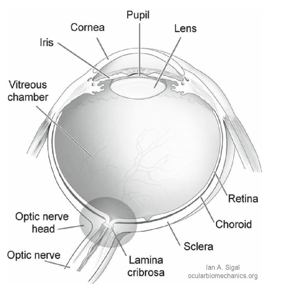
Schematic cross-section through a human eye. Light enters the eye through the cornea, passes through the pupil, lens and vitreous humour and strikes the retina, where it is absorbed. Retinal nerve fibers transmit visual information to the brain. These fibers converge at the optic nerve head region, exit the eye through the scleral canal, and form the optic nerve. The lamina cribrosa is a porous structure spanning the scleral canal. The vitreous chamber is filled with the vitreous humor, which exerts a pressure, the intraocular pressure, on the surface of the retina. [Sigal et al. Biomech Model Mechanobiol, 8(2):85-98, Apr 2009] (adapted from an illustration from NIH) |
Goals
The objective of the Laboratory of Ocular Biomechanics is to study the eye as a biomechanical structure. More specifically our work is aimed at identifying the causes of glaucoma, with the ultimate intention of finding a way to prevent vision loss.